Wednesday, October 27, 2010
Scribepost oct. 27 by Danyal
- Bacteria
- endemic/diarrheal
- transmission through contaminated food or water
- targets intestines epithelial cells
- treated with antibiotics
- prevented by purified water, sewage treatment, and proper cooking
- 100,000-200,00 deaths/year world wide

This is a picture of
the cholera virus
These were the notes we took today in addition to a hand out he gave us about bacteria.
Bacteria
- unicellular prokaryotes [lack a nucleus] or other membrane bound organelles
- Contain a cell wall
- Bacteria are separated into 2 kingdoms eubacteria[common bacteria] and archaebacteria
Eubacteria- exists almost everywhere has cell wall made of peptidoglycan.[a carbohydrate]
This is a picture of Eubacteria.

Here is a picture of of Archaebacteria.

Shapes of bacteria
- Bacilli [rod shaped] picture

2. cocci [spherical shaped] picture

3. sprillum [spiral] and corkscew shaped] picture

METABOLISM[ENERGY]
- heterotrophs- obtain energy from organic molecules
- Autotrophs- make their own food from in organic molecules
- Phototroph- make own enrgy from light
- chemotroph- lives of energy from chemicals
- saprophytic- live of dead or decaying organic matter[decomposer]
METABOLISM[RESPIRATION]
- obligate aerobes- organisms that require a constant supply of oxygen
- obligate anaerobes- organisms that do not require oxygen and some die with oxygen
- facultative anaerobes- organisms that can survive with or without oxygen
At the end of class we watched a video on bacteria.
END
Monday, October 25, 2010
Scibepost for October 25, 2010 by Jiyoon
Today, Mr. Paek reminded us that the Cancer Paper is due tomorrow. We have to make sure to turn it in to www.turnitin.com and ALSO bring in a hard copy to turn in for class. He also told us that it's a good idea to have quotes in our papers and to make sure to have the bibliography within your paper. Don't forget to read 20.1 for tomorrow and 20.2 for Wednesday, you don't have to take notes.
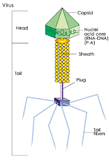
- non-cellular particles (no organelles)
- genetic material (DNA/RNA)
- Protein (outer coat or capsid)


- cancer paper
- reading 20.1
- try to finish the lab
Thursday, October 14, 2010
Scribepost- October 14th, 2010- Fox :D


 Picture Copyright goes to Yulia B. DO NOT TAKE WITHOUT PERMISSION. thank you. (just kidding, do whatever you want with it)
Wednesday, October 13, 2010
Scibepost for October 13, 2010 by Amreen M(=
First, he explained that the reason we don't have a bunch of big cells in our body is because with small cells, theres more surface area in terms of volume so its easier for gases to get exchanged.
 Mr. Paek explained to us that interphase, which are the three big parts of this cycle, takes much longer then metaphase, which are the four small parts. Interphase has three parts: the G Phase, S Phase, and the G2Phase, and it basically is preparing the cell to divide. During interphase, the individual chromosomes cannot be distinguished and appears as a dark mass of material called chromatin.
Interphase- Each chromosome is replicated, consisting of two identical "sister" chromatids; the DNA of each chromosome replicates at the end of this stage.
After he explained Interphase for a couple minutes, he went on to mitosis, which has four stages. Phrophase, Metaphase, Anaphase, and Telephase. Mr. Paek said that when he was learning the cell cycle in school, he used an anagram called IPMAT, which stands for Interphase, Prophase, Metaphase, Anaphase, and Telephase. These stages are also in order.
 This is a picture of a cell in the prophase stage.You can tell its in the phrophase stage because the membrane has holes in is and is beginning to break.
 Anaphase-The paired chromosomes split into individual chromosomes and are moved apart.


We also briefly talked about Cytokinises, which takes place after Telophase. Cytokinesis is when the cytoplasm pinches in half and each daughter cell has duplicate chromosomes. Here is picture summing up everything:

After we were finished taking notes, Mr. Paek let us watch the Last Lecture for the last two minutes of class. Our homework is to read, highlight, and complete page 40-43 in our Unit Packet. These pages are our Mitosis Pre-Lab for the lab that we will be doing tomorrow
The next scriber is..MALIHA!=D
Monday, October 11, 2010
Scribe post for October 11th, 2010 by Junsup Lee
Independent variable is on X-axis
dependent variable on the Y-axis
3 definition that will help you during lab tomorrow

-Crenated is word for animals
-plasmolysed is word for plants
-Plasolysed: meaning when cell shrinks in hypertonic solution
2. Isotonic solution: Cell shape dosen't change. Same ratio of water coming out and same amount going in. The middle one of the picture above. Its just regular size of blood cell.
3.Hyponic solution : more amount exist insides so it trieds to go out so the cell expands. Third picture above the blood cell got bigger. More thing comes out and Less thing comes in.
makes sure you know all these definitions above
OUR HOMEWORK TODAY is to finish the Enzyme lab it is page 24 to 32 of your Unit packet
Sunday, October 10, 2010
Scribe Post for October 8th, 2010 by Richard P.
The Pre-lab was due for homework today, so if you were unable to complete it, have it ready for Monday, October 11.
Today, we did a lab involving Enzymes. The Lab is detailed on Pages 24 through 32 in your Unit Packet.
For those of you who don't know, Enzymes are proteins that speed up chemical reactions in cells. Almost all chemical reactions that occur in living organisms (For example, digestion) are catalyzed (sped up) by enzymes. Many factors in the environment of a cell affect the action of an enzyme. For example, enzymes work best at certain pH values and at certain temperatures.
Catalase is an Enzyme found in blood and other cells. It speeds up the breakdown of hydrogen peroxide (the substrate) into water and oxygen. If left to build up within cells, hydrogen peroxide is toxic.


The Materials we needed for the lab were:
1. Catalase (enzyme)
2. 6% hydrogen peroxide solution (substrate)
3. water
4. dropper pipette
5. test tubes
6. 25-mL graduated cylinder
7. metric ruler
8. glass-marking pencil
9. ice bath
10. thermometers
11. warm water bath
12. safety glasses
All materials were provided for us.
For the Lab, we had pre-assigned groups of 4 people each. Specific temperatures were assigned to each group.
In the Enzyme Lab we did,we tried to determine the effect of different temperatures on enzyme activity. In the lab, we subjected the Catalase Enzyme to different temperatures. We then added hydrogen peroxide to the test tube, and we then measured how much activity occurs by measuring the height of the oxygen bubble column in the test tube. The higher the bubble column, the more enzyme activity there was.
The Lab was divided into two sections: Part A: Observe the Catalase Reaction and Part B: How does Temperature affect Enzyme Activity?
Part A: Observe the Catalase Reaction Steps:
1. Put on your gloves and safety goggles
2. With the red marking pencil, label 3 test tubes at the 1 cm and 5 cm levels (Measure from bottom of test tubes.)
3. Fill test tube #1 to the first mark with catalase (enzyme). Then, fill test tube #1 to the second mark with hydrogen peroxide (substrate). Swirl to mix them together. Then wait 20 seconds for the bubbling to occur.
4. Measure the height of the bubble column in mm, then record the data in the first table that corresponds to the lab in the Unit Packet.
5. Next, fill test tube #2 to the first mark with water. the, fill test tube #2 to the second mark with hydrogen peroxide (substrate). Swirl to mix. Wait 20 seconds for bubbling to occur.
6. Measure the height of the bubble column in mm, and record in the first table.
7. Lastly, fill test tube #3 to the first mark with catalase. next, fill Test tube #3 to the second mark with sucrose solution (a different substrate). Swirl to mix. Wait 20 seconds for bubbling to occur.
8. Measure the height of the bubble column in mm, and record it in Table 1 of your Unit Packet.
We then had to answer questions about Part A.
Part B: How does Temperature affect Enzyme Activity? Steps:
1. Formulate a Hypothesis
2. Test your hypothesis. Outline a brief procedure for your experiment.
3. Set up your warm or hot water, or ice bath to achieve your desired temperature.
4. With the red marking pencil, label a new test tube at the 1 cm and 5 cm levels.
5. Fill your test tube to the first mark with catalase enzyme.
6. Place your test tube into the water bath of your assigned temperature for 10 minutes. Monitor the temperature of the catalase until it reaches the desired temperature.
7. After 10 minutes, use your thermometer to record thee actual temperature of the catalase. Remove your test tube from the water bath, and add hydrogen peroxide (substrate) to the second line. Swirl to mix. Wait 20 seconds for bubbling to occur.
8. Measure the height of the bubble column in mm. Make a table of results and record the data in it.
9. Repeat the experiment one more time.
10. Determine your average temperature (Celsius). Record your data on the sheet with the class
data.
For homework, we have to start on the analysis and interpretation questions in our Unit Packet starting on page 27. We do not need to finish it, but simply start it.
- Richard P.
Thursday, October 7, 2010
Yet Another Scribepost by Josh B, on Enzymes, p4sts2010
Don't misunderestimate my note taking power, by the way. (Word from George Bush).
Homework
-Next, Mr. Paek decided to be insane and put Potassium Iodide (KI) into a large cylinder that had some Hydrogen Peroxide (H2O2) in it. It exploded... in a huge column of condensed steam that looked like a solid. It just kept foaming up and breaking off over the top of the cylinder. This picture isn't from class, but sort of portrays the reaction.
 This was really cool, and if you missed it, then that sucks.
-Mr. Paek also decided to use a new system of punishment for talkative people. If you are talking too much, he will take you to the front of the room and make you put your finger in Hydrochloric Acid for several minutes. Just kidding (or am I...). Actually, if you are talking, your name will be written on the board, and a point will be taken off your grade. Another offence, and that number is doubled. For more information, talk to Jake C.

I like money

THE VICTIM FOR TOMORROW WILL BE CHOSEN BY THE FORCE OF GOD!
GOD CHOOSES... Richard
Wednesday, October 6, 2010
Wednsday, 10/6/10
- A tied-off cellophane dialysis tube is filled with a starch solution and then a glucose solution to represent a membrane full of fluid.
- The filled tube of starch and glucose is submerged into a glass of water and iodine, where it will sit for about 15 min. (During this time, we took the Organelle Quiz.)
- Upon returning, the solutions in the tube and the glass should show some results. If the iodine water in the glass has turned blue, that means there is starch in the water. Then, a piece of Tes-Tape is dipped into the iodine water, and if the tip turns green or yellow, then there is glucose in the water. Meanwhile, if the tube full of starch and glucose solution has turned purple, then there is iodine inside the tube.
- For our group's lab test, the membrane tube turned purple, the iodine water solution stayed orange and didn't change color, but the piece of Tes-Tape turned blue instead of green or yellow.

Overall, it can be concluded that the glucose passed through the tube and went into the water surrounding it, the iodine in the water passed through the tube and went inside, the starch never went anywhere and stayed in the tube, and the water passed throughout freely. This is explained by the fact that the cellophane tube is full of holes that can allow individual molecules to pass through one by one through each tiny hole. This is what allowed the small iodine and glucose molecules to pass slowly through the barrier of the tube, but starch is made up of larger molecules made from glucose, which is why starch was unable to transfer through the tube at all.
As for our own experiment, however, the tape turning blue rather than green or yellow was probably a small fault in our preparation.
BTW, who's the next scriber? :P
Tuesday, October 5, 2010
Tuesday-Carly
-nucleus: DNA
-Nucleolus: the assembly of Ribosomes; Proteins
-Golgi Apparatus: produced in Rough ER; appears as a stack of pancakes.
-Lysosomes- break down lipids, proteins, and carbohydrates.
-Vaculoes: stores materials
-Mitochondria: power house of the cell; where the cell gets energy
-cytoskeleton: maintains shape and provides movement
-chloroplast- takes energy from the sun
-cell membrane: 2 layers of fat ( lipid bilayer), regulates what goes in and out of the cell
After this everbody took notes on...
Monday 10.4.10
10.4.2010 Today in Biology, we worked on two worksheets. On one we filled out chart on the many organelles. It was kind of like the one below. The columns were; the organelles function, which cell its found in, an analogy to a city and a sketch of the organelle. example; | Vacuoles | both | stores materials | (picture) | Storage Places (P.O.D.) | The second worksheet, we had to draw a cell and label it.
Organelles that Store Clean Up and Support -Vacuoles - Has vesicles that store and move cells to and from cells -Lysosomes - Clean up crew - Remove waste -Cytoskeleton - Microfilaments; support the cell - Microtubes; important with cell division Organelles That Build Proteins -Ribosomes - Makes proteins -Endoplasmic Reticulum - Have smooth and rough ET -Golgi Apparatus - Gets proteins to the right places Organelles that Capture and Release Energy -Chloroplasts - Same a solar plants - Contain the green pigment chlorophyll -Mitocandria - Have their own small DNA Cellular Boundaries -Cell Wall - Animal cells do not have cell walls - Nearly all of trees and stems are made up of cell wall material -Cell Membrane - Has a lipid bilayer - Allows some things to pass through and other things not the next scriber will be...... Carly! |
Monday, October 4, 2010
7.2 Texter---Grace

- Cytoplasm- Portion of the cell outside the Nucleus
- Organelles-Specialized structure that performs important cellular functions within an eukaryotic Cell
- Vacuoles- Structures in a cell that stores diffrent materials
- Lysosomes- Small organelles filled with enzymes
- Cytoskeleton- A network of protein filaments
- Microfilaments-threadlike structures made up of a protein called actin
- Microtubules- hollow structures made up of protein called tubulins
- Centrioles- Organelles that help organize cell division
- Ribosomes- small particles of RNA and protein found throughout the cytoplasm in all cells
- Endolplasmic Reticulum- An internal membrane system
- Golgi Apparatus- Organelles that modifies, sorts, and packages protein and other materials for storage in a cell or release outside the cell
- Chloroplast- Organelles that capture energy from the sunlight and converts it into food
- Mitochondria- Organelles that convert solar energy stored in food into copounds that are more convenient for the cell to use
- Cell Wall- a strong supporting layer around the membrane of a cell
- Lipid Bilayer-flexible double layered sheet that makes up the cell membrane and forms a barrier between the cell and its surroundings
- Selectively Permeable- Substances that can pass across cell membrane and other can't
- The nucleus contains most of the cells DNA and controls the functions of the cell
- Chromosomes contain the cells genetic information
- Nuclei are contained in the nucleolus
- Proteins are assembled on ribosomes
- Proteins created on the Endoplasmic Rectum includes ones that will be released, membrane proteins, and proteins for lysosomes and other specific locations in a cell
- Most cell walls have enough pores to allow water, oxygen, carbon Dioxide, and other substances pass through
- The cell membrane chooses what goes in and out of the cell as well as it also protects and supports the cell
- Most of the cell membrane is protein molecules
Sunday, October 3, 2010
10/3/2010
